6515 Main Street
Trumball, CT 06611

Articles

Visual field testing is an important part of most standard comprehensive eye exams. Also sometimes known as perimetry testing, Visual field testing is a method to measure the entire scope of vision of an individual, including their peripheral/side vision.
The importance of visual field testing
Visual field testing is one of the most effective diagnostic treatments in the detection of glaucoma. This is because when patients are affected by glaucoma, it is usually the peripheral vision that is affected by their condition first. However, it can also be used to detect central or peripheral retinal diseases, eyelid conditions such as drooping, optic nerve damage and conditions that affect the visual pathways from the optic nerve to the area of the brain where this information is processed into vision.
Visual field testing is also an important part of monitoring for people who are considered to be at risk for vision loss from disease and other problems, including those who have been diagnosed with the following:
Multiple sclerosis
Hyperthyroidism
Pituitary gland disorders
Central nervous system problems (such as a tumor that may be pressing on the brain)
Stroke
Diabetes
High blood pressure
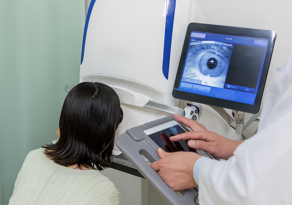
Optomap is an innovative new technology that gives eye doctors the ability to perform ultra-wide retinal imaging that is far superior to what can currently be achieved using conventional retinal imaging options. In contrast to conventional retinal imaging, Optomap captures at least 50% more of the retina in a single capture, and with Optomap’s multi-capture function, up to 97% of the retina can be viewed. This gives eye care professionals greater opportunity to monitor the health and condition of patient vision.
Why is Optomap important?
Optomap is another great preventative eyecare technology tool. By allowing your eye doctor to have a comprehensive view of your retina, they will be able to detect any developing eye diseases early on, before they have a detrimental impact on your vision and day to day life. Not only can Optomap detect eye conditions such as retinal holes, retinal detachment, macular degeneration and diabetic retinopathy, but it can also be used to identify some general health conditions such as cardiovascular disease, stroke and cancer.

Presbyopia is a natural and inevitable part of aging that affects the eyes' ability to focus on close objects. Unlike other vision conditions, presbyopia isn’t caused by the shape of the eye or structural abnormalities but is instead a result of the eye’s lens losing its flexibility over time.
What is Presbyopia?
Presbyopia is a refractive error, meaning it affects how the eyes bend (or refract) light to focus it on the retina. Unlike nearsightedness (myopia) or farsightedness (hyperopia), which are caused by the shape of the eyeball or cornea, presbyopia is due to the aging of the eye's lens. Over time, the lens becomes less flexible, making it difficult to focus on nearby objects. Presbyopia isn’t a disease or an abnormality—it’s simply a natural consequence of the aging process.
What Causes Presbyopia?
The lens inside the eye changes shape to focus light on the retina, allowing us to see clearly at varying distances. When viewing nearby objects, the lens thickens and curves to increase its refractive power.
As we age, however:
Lens Stiffening: The lens loses elasticity, making it harder to bend and adjust for near vision.
Weakened Ciliary Muscles: The muscles that help change the shape of the lens may lose their strength over time, further reducing the eye’s ability to focus on close objects.
Structural Changes: The lens also grows thicker and less transparent with age, which contributes to reduced focusing ability.
These changes are gradual, typically starting in a person’s 30s and becoming noticeable by their early to mid-40s.
InfantSEE
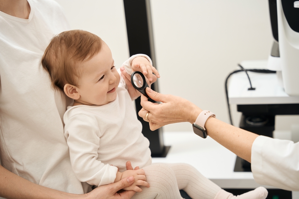
Your baby’s vision develops quickly during the first year of life and plays an important role in how they learn, move, and interact with the world. Even when a baby’s eyes appear healthy, certain vision or eye health concerns may not be obvious without a comprehensive infant eye assessment. InfantSEE is designed to help identify potential concerns early, giving parents peace of mind and helping children start life with the clearest, healthiest vision possible.
What Is InfantSEE?
InfantSEE is a program that helps parents access an early, comprehensive eye and vision assessment for babies between 6 and 12 months of age. It is designed to support healthy visual development by giving us an opportunity to check for early signs of vision or eye health concerns during a critical stage of growth.
The assessment is provided at no cost, making early eye care more accessible for families who want to be proactive about their baby’s vision.
The Importance of Early Vision Care
Many parents are surprised to learn that babies can have vision or eye health issues even before they are old enough to talk or read an eye chart. An infant may still track faces, reach for toys, or appear visually engaged while having an underlying concern that needs attention.
Early eye care can help detect signs of:
Eye alignment problems, such as crossed or turned eyes
Significant nearsightedness, farsightedness, or astigmatism
Eye movement or focusing concerns
Amblyopia risk factors, often called lazy eye
Signs of eye health conditions that may need monitoring or treatment
Finding these concerns early can make treatment more effective and may help support long-term visual development.

Myopia is a common vision problem affecting a large number of individuals worldwide. It occurs when the eye is unable to focus properly on distant objects, resulting in blurred vision. Myopia can develop during childhood and progress into adulthood if left unmanaged.
What Causes Myopia to Develop?
Myopia can be caused by a combination of genetic and environmental factors. If one or both parents have myopia, there is a higher chance that their children will develop the condition. Additionally, excessive screen time, lack of outdoor activities, and prolonged near-work activities such as reading or using electronic devices can contribute to the development of myopia. These lifestyle factors can lead to the elongation of the eyeball, resulting in the inability to focus on distant objects.
The Importance of Myopia Management
Managing myopia is crucial to prevent its progression and associated complications. High levels of myopia can increase the risk of developing eye conditions such as glaucoma, cataracts, and retinal detachment.
Additionally, severe myopia can lead to vision loss and significantly impact the quality of life. Early intervention and proper management of myopia can help slow down its progression and reduce the risk of these complications.

Our eyes are extremely delicate, yet they can be subjected to harsh conditions and other environmental factors that affect their health. One of the problems that can affect our eyes is an accumulation of dirt, debris and bacteria on the eyelids. This can cause a range of issues, including stopping tear film from reaching the eyes and being properly dispersed over their surface – which is necessary to keep them healthy and comfortable. Fortunately, a new solution called Blephex® can help.
What is Blephex®?
Blephex® is a handheld electro-mechanical device that is applied to the margins of the eyelids with the purpose of cleaning them and improving the effectiveness with which tear film flows onto the surface of the eyes.
Blephex® has a disposable, surgical-grade sponge tip which rapidly oscillates to create a cleaning action. Before the sponge tip is placed onto the eyes, it is soaked in a gentle exfoliating solution. This solution provides soft abrasion to help remove dead skin cells and debris that could be irritating the eyes and interrupting tear film progression. The Blephex® device is manually applied to the eyes and moved gently across the eyelids, with the entire, painless process taking approximately 6 to 8 minutes per eye. A different sponge is used on each eye, ensuring that no bacteria is passed between them. After the procedure, patients are given instructions on how to maintain the cleanliness of their eyelids with daily/nightly eyelid hygiene at home.
Most patients experience a significant improvement in tear film production and dispersal, and a reduction in unpleasant symptoms that they may have been experiencing within 48 hours of their treatment. While a single treatment is normally enough to produce excellent results, many patients are advised to have Blephex® every 4-6 months.

Millions of people suffer from eye and vision-related problems. For some, the solution is as simple as wearing prescription eyeglasses or contact lenses. However, others face challenges that aren’t as easy to rectify. That’s where Lumenis comes into play.
As a world leader in minimally invasive clinical solutions for both ophthalmology and aesthetic markets, it’s making a significant difference. Not only does Lumenis develop advanced energy-based technologies, but it also commercializes them.
The company continues to find innovative solutions. However, it’s already provided the fields of ophthalmology and aesthetics with remarkable technologies. OptiLight is just one example.
Eye and Vision-Related Solution
The company invented the first and only technology to treat individuals for dry eye disease caused by MGD. Not only is it approved by the Food and Drug Administration (FDA), but also patented. Optimal Pulse Technology (OPT) makes it easier to successfully manage this particular eye disease. Overall, OptiLight is safe, precise, comfortable, and effective.
Managing Symptoms
According to recent statistics, roughly 22% of the U.S. population suffers from dry eye disease along with MGD. That combination leads to an array of uncomfortable symptoms.
Dry eye occurs when a person’s eyes don’t produce adequate tears to keep them lubricated or when tears don’t work the way they should. MGD is a condition that affects the small glands in the eyelid responsible for making the oil layer for tears.
Having to deal with one of these problems is bad enough. However, living with both can prove debilitating for some people. Thanks to OptiLight, people with the two conditions get much-needed relief. Here are the ways this advanced technology helps:
Reduces inflammatory mediators, which, thereby, prevents inflammation
Improves tear breakup time, which, in response, decreases osmolarity
Alleviates abnormal blood vessels that commonly cause inflammation
Restores meibomian glands so they function properly
Decreases demodex mites that cause infection and the accumulation of bacteria on the eyelids

Telemedicine makes it easier to connect with your optometrist when an in-office visit is not necessary or not possible. A virtual eye care appointment allows you to discuss certain eye health concerns, follow-up needs, and symptoms through a secure video visit from home, work, or another private location.
All you need is a smartphone, tablet, or computer with a camera, microphone, and internet connection. The team will provide instructions before your appointment so you know what to expect and how to join your virtual visit.
How A Virtual Eye Care Visit Works
To schedule a telemedicine appointment, call our office or request an appointment online. Before confirming your visit, we may ask a few questions about your symptoms, medical history, and current eye care concerns. This helps us determine whether a virtual visit is appropriate or if you should be seen in person.
During your appointment, your optometrist will review your concerns, ask about your symptoms, and may guide you through simple observations or questions to better understand what is happening. In some cases, photos of the eye or eyelid may be helpful if they are clear and taken safely.
Neuro-Optometric Rehabilitation
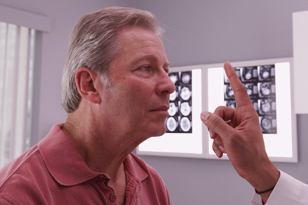
When most people think about vision problems, they picture issues with the eyes themselves. However, many visual difficulties actually begin in the brain. After a concussion, stroke, or other brain injury, the connection between the eyes and brain can be disrupted - causing symptoms that affect balance, reading, focus, and even daily comfort.
Brain Injuries and Vision
Your visual system is complex - about 70% of the brain is involved in vision processing. After a traumatic brain injury (TBI), concussion, or neurological event such as a stroke, the pathways that coordinate eye movement, focus, and perception can become impaired.
This can result in symptoms such as:
Blurred or double vision
Eye strain and headaches
Difficulty reading or focusing
Dizziness or balance problems
Light sensitivity
Problems with depth perception
These visual issues often persist long after other symptoms of a brain injury have improved, making specialized vision care essential for recovery.
What Is Neuro-Optometric Rehabilitation?
Neuro-optometric rehabilitation is a specialized form of vision therapy designed to retrain how the brain and eyes work together. Unlike traditional eye care that focuses on eyesight alone, this therapy targets the neurological processes behind vision. Our doctor evaluates how the eyes and brain communicate and creates a personalized treatment plan to rebuild these visual pathways. The goal is to restore comfort, coordination, and clarity - improving overall visual function and quality of life.

Strabismus is a relatively common condition, affecting about 2-4% of the population worldwide. It is one of the most frequent eye disorders seen in children, though it can also develop in adults due to conditions like stroke or trauma. In the United States alone, it is estimated that around 12 million people are affected by strabismus, with varying degrees of severity. Early diagnosis and treatment are essential, as untreated strabismus in children can significantly impair visual development.
What is Strabismus?
Strabismus, commonly referred to as crossed eyes, is a condition in which the eyes do not properly align with each other when looking at an object. One eye may turn in, out, up, or down, while the other eye focuses correctly. This misalignment can be constant or intermittent and may affect one or both eyes. Strabismus occurs when the muscles controlling eye movement do not work together correctly, leading to difficulty in focusing both eyes on a single point. This condition can lead to visual issues such as double vision and, in some cases, can result in amblyopia (lazy eye) if left untreated.
How Does Strabismus Develop?
Strabismus develops when there is a disruption in the coordination of the muscles that control eye movement, leading to a misalignment of the eyes. This disruption can result from various causes, including genetic factors, neuromuscular conditions, or problems with the nerves that transmit signals to the eye muscles. In some cases, it may be present at birth, while in others, it can develop later in childhood or adulthood due to conditions such as cerebral palsy, Down syndrome, or even trauma to the eye or brain.
Signs and Symptoms of Strabismus
The primary symptom of strabismus is the visible misalignment of the eyes. However, there are other signs and symptoms that may indicate the presence of this condition:
Blurred or double vision
Headaches or eye strain
Difficulty with depth perception and spatial awareness
Reduced visual acuity in one eye
Frequent eye rubbing or squinting
Tilting or turning the head to compensate for the misalignment

Services
Learn More
Keep
In Touch
Ready to Begin
Your Healing Journey?
Take the next step toward better vision, deeper wellness, and lasting transformation.
Managed and Designed by:
